
Inside a Bled AVM: What Dr. Vikas Gupta Shared at NVISICON 2026?
There is a particular kind of quiet that settles over a conference hall when a speaker puts up a scan of a 22-year-old with a headache that turned out to be a bleed in the brain. That was how Dr. Vikas Gupta opened his talk, “Bled AVM: Simultaneous Endovascular Embolisation & Excision,” at NVISICON 2026, the Summer Colloquium of the Neurovascular Interventions Society of Indraprastha (NVISI), themed “Basics and Precision in Stroke Neurointervention.”
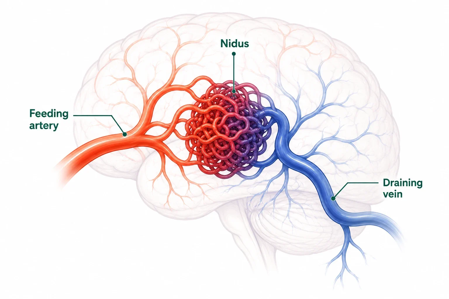
What Was the Talk at NVISICON 2026 About?
Dr. Gupta’s session centred on one specific, high-stakes question: when a brain AVM has already bled, does it make more sense to treat it in two separate procedures, or in one combined sitting?
Rather than presenting theory alone, he walked the room through a series of real patients, each with a different AVM location, a different bleed pattern, and a different reason the case had stayed with him.
The talk opened with a map of how AVM treatment approaches vary across India:
- Radiosurgery-first: The preferred approach in some centres
- Surgery-only or embolisation-only: Used as standalone techniques in other centres
- Multimodality approach: Combining more than one technique, the approach favoured in Delhi
This regional variation set up the real argument of the talk.
Why Combine Embolisation with Surgical Excision?
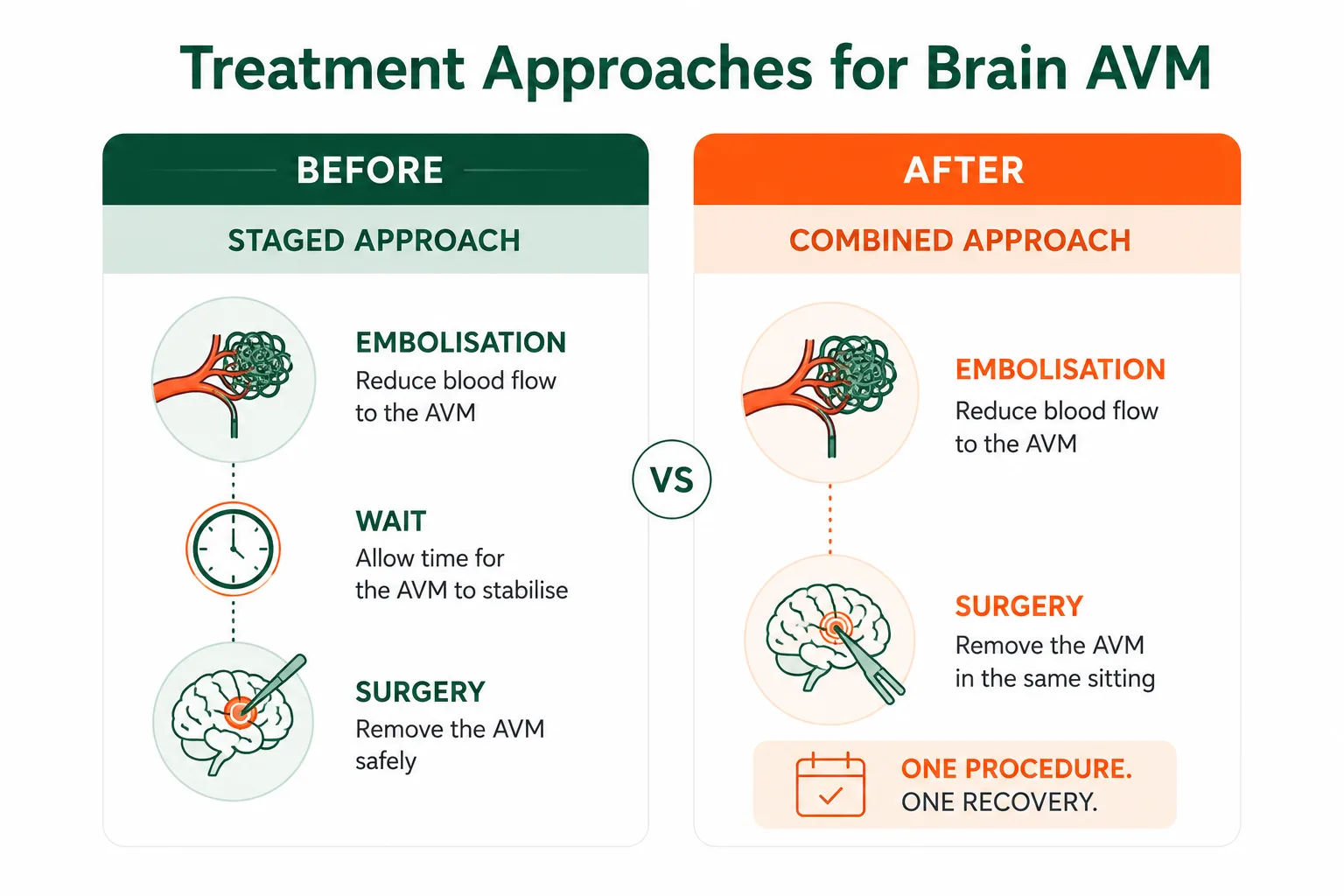
Combining endovascular embolisation with microsurgical excision in the same setting can reduce blood loss during surgery and shorten the time a ruptured AVM is left only partly treated.
Embolisation works by threading a catheter through the blood vessels to plug the AVM’s feeding arteries with a glue-like agent, cutting off much of its blood supply. Excision is the surgical removal of what remains. Done separately, there can be a gap of days between the two, a gap Dr. Gupta’s cases suggested is worth closing wherever it’s safe to do so.
The AVM Cases That Carried the Talk
Rather than presenting theory alone, Dr. Gupta walked the room through six real patients, each with a different AVM location, a different bleed pattern (or, in one case, no bleed at all), and a different reason the case had stayed with him.
Case 1: 22-year-old man, headache and vomiting
- Scans showed a cone-shaped tangle of vessels, classic AVM angio-architecture, feeding a deep bleed
- Angiogram, Nidus embolisation, Microsurgical excision (same admission)
- Post-op scans: no residual AVM, a recovering brain
Case 2: 42-year-old man, headache from a bled AVM
- DSA showed the full run: right ICA injection, nidus, draining vein
- Obliteration with a liquid embolic agent
- Surgery
Case 3: 11-year-old boy, seizures with brief loss of consciousness
- Sulcal AVM
- Embolisation
- Obliteration
- Clean post-operative course
Case 4: Prem Singh, 44-year-old man, trigeminal neuralgia (no bleed, the AVM announced itself differently)
- Sudden, severe facial pain, worse while sleeping, traced to a posterior fossa AVM pressing on the nerve
- MRI/MRA, Embolisation with Onyx, Further glue injections and obliteration runs, Surgery (sitting position)
- Outcome: pain-free, no residual disease on control angiogram
Case 5: 30-year-old woman, brought in unconscious
- Left-sided weakness, unequal pupils (anisocoria)
- CT and CT angiography, AVM localised, Combined treatment
- Outcome: full orientation regained by day 14
Case 6: 31-year-old woman
- Embolisation, Surgical excision (later)
- Outcome: marked improvement in limb power
Dr. Gupta was candid that none of these were simple cases; the real value was in showing, angiogram by angiogram, how the decision to combine techniques was made each time.
What Did The Delegates Take Away?
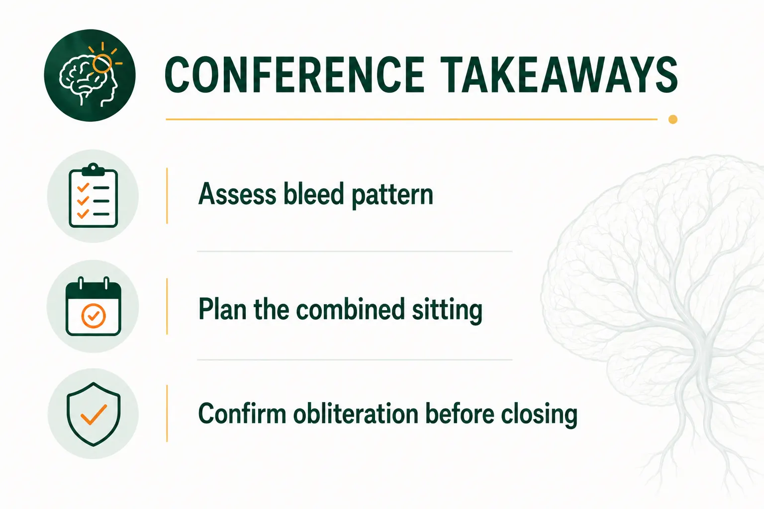
For the neurointerventionists and neurosurgeons in the room, the takeaway wasn’t a single formula; AVMs vary too much in size, location, and drainage pattern for that.
It was closer to a working principle: when a bled AVM is amenable to it, treating the feeding vessels and the nidus in one coordinated sitting, rather than two spaced-out ones, can shorten the window of vulnerability for the patient.
Dr. Gupta grounded this in the Spetzler-Martin grading system that guides much of AVM decision-making, while acknowledging that grading is only the starting point; the angiogram in front of you still has the final say.
For patients and families reading this rather than sitting in a conference hall, the practical point is simpler: a bled AVM is treatable, and the “how”, embolisation, craniotomy and excision, or both together, depends on where the AVM sits and how it’s behaving, which is exactly the kind of detail a proper angiogram and consultation is meant to work out.
If you or a family member has been told there’s an AVM involved in a brain bleed, a conversation with a neurointerventional specialist about whether a combined approach applies to your case is a reasonable next step.
Frequently Asked Questions
What is the advantage of combining embolisation and surgery for a bled AVM? expand_more
Combining the two reduces blood loss during surgery and shortens the time a ruptured AVM is left only partly treated, since the feeding vessels are blocked before the surgical team removes what remains, rather than leaving a gap of days between procedures.
What should I do if I or a family member has been told an AVM caused a brain bleed? expand_more
A prompt consultation with the best neurosurgeon is advised. Imaging (MRI/CT/DSA) and AVM grading will determine whether embolisation, surgery, radiosurgery, or a combined approach is appropriate for that specific case.
Who is the best neurosurgeon in Delhi NCR for brain AVM treatment? expand_more
Dr. Vikas Gupta is a Senior Consultant Neurosurgeon and Neurointerventional Specialist with over 30 years of experience and has managed complex and ruptured AVM cases. Patients are advised to review a specialist's case experience, hospital infrastructure, and case-specific fit before deciding.
How can I book an appointment with Dr. Vikas Gupta? expand_more
You can book an appointment with Dr. Vikas Gupta by contacting us directly, using our online appointment booking facilities, available on this website.
How can I book an appointment at Kailash Deepak Hospital? expand_more
You can book an appointment at Kailash Deepak Hospital by calling the hospital reception directly or using the online appointment booking facility on the hospital's website.

 WhatsApp