
Bilateral Subdural Haematoma Treatment Without Open Brain Surgery
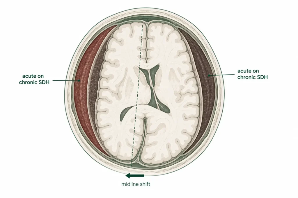
Headache. Confusion. Weakness. He Was 80, and Blood Was Pressing on Both Sides of His Brain.
At a Glance
| Patient | 80-year-old male |
| Complaint | Headache, altered sensorium, generalised weakness |
| Diagnosis | Bilateral acute on chronic subdural haematoma, left greater than right |
| Procedure | Cerebral DSA followed by bilateral Middle Meningeal Artery embolisation |
| Outcome | Reduction in brain swelling and midline shift confirmed on post-operative CT; clinical improvement noted |
An 80-year-old man came in with headache, confusion, and weakness across his body. He was not fully alert. His responses were slow. Something was pressing on his brain, and it was pressing on both sides.
What followed was a careful clinical decision: how to treat bilateral brain bleeds in an 80-year-old without the risks of open surgery.
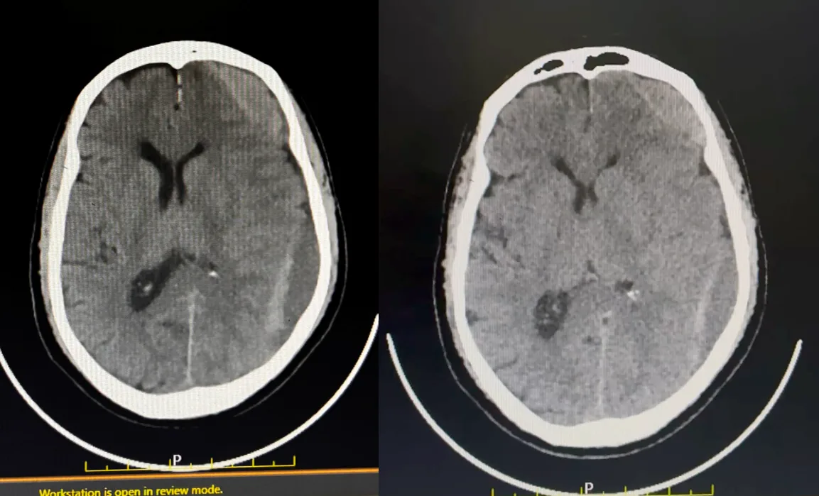
CT scan showing large left-sided subdural haematoma with mixed density,
indicating acute bleeding on top of a chronic collection.
Reach Out for Expert Care
What Did The Scan Show?
Imaging confirmed blood collections on both sides of the brain, old bleeds with fresh bleeding on top, the left side significantly larger and causing the brain to shift.
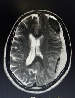
MRI brain showing bilateral subdural haematoma collections along both cerebral convexities,
left greater than right.
The CT scan showed a large extra-axial mixed-density collection along the left fronto-temporo-parietal region, a technical way of describing a significant blood collection sitting over a wide area of the left side of the brain.
The mixed density, both old and new blood visible together, indicated an acute on chronic subdural haematoma: a chronic collection that had been building over time, with a fresh bleed layered on top of it.
The MRI confirmed the full picture: bilateral concavo-convex collections along both cerebral convexities, left greater than right. The brain was being compressed from both sides, with the left-sided collection large enough to cause a shift in the brain’s midline, a sign of significant mass effect that required urgent treatment.
This explained everything: the headache from rising pressure, the confusion from compressed brain tissue, and the generalised weakness from disrupted neurological function.
The Challenge
Bilateral SDH in an elderly patient is a high-stakes situation. Treating one side is complex enough; treating both sides, in an 80-year-old without open surgery requires precise planning and endovascular expertise.
In acute on chronic SDH, the haematoma has two components: the old, organised collection that has been building over time, and the fresh bleed on top of it. Simply draining the blood addresses the immediate crisis, but does not stop the Middle Meningeal Artery from continuing to supply the outer membrane, which drives reaccumulation.
In an 80-year-old patient, open bilateral craniotomy or burr hole drainage carries a substantially elevated risk. The endovascular approach, Middle Meningeal Artery embolisation, targets the root cause of the problem by cutting off the blood supply feeding both haematoma membranes, from within the blood vessels themselves, without a single incision on the skull.
Cerebral Digital Subtraction Angiography and Bilateral Middle Meningeal Artery Embolisation Procedure: What Was Done?
Dr. Vikas Gupta performed Cerebral Digital Subtraction Angiography followed by bilateral Middle Meningeal Artery embolisation, treating both sides in a single minimally invasive procedure.
A catheter was carefully navigated through the blood vessels under continuous image guidance. A cerebral DSA, a detailed angiogram of the brain’s blood supply, was performed first to map the middle meningeal arteries on both sides and confirm their supply to the haematoma membranes.
Both left and right Middle Meningeal Arteries were then selectively embolised, small particles delivered precisely through the catheter to block the blood supply feeding the haematoma membranes on each side. With both feeding arteries blocked, the membranes driving the reaccumulation of blood on both sides were deprived of their supply, allowing the body to gradually reabsorb the collections over the weeks that follow.
The entire procedure was performed without any incision on the skull.
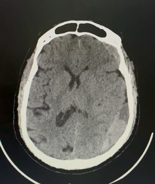
Post-operative CT scan confirming reduction in brain swelling and mass effect,
with decreased midline shift, evidence of early response to embolisation.
Outcome
Post-operative CT confirmed meaningful early results: the brain swelling and mass effect had reduced, and the midline shift, the clearest indicator of how much pressure the brain had been under, had decreased. These imaging changes confirmed that the embolisation had achieved its goal of interrupting the blood supply driving the haematoma.
Clinically, the patient showed improvement, clearer sensorium, reduced headache, and improvement in generalised weakness. In bilateral chronic SDH cases, full resolution of the haematoma is a gradual process as the body reabsorbs the old blood collections over weeks following successful embolisation. The imaging and clinical trajectory at the time of review were both positive.
Why Does This Case Matter?
Bilateral subdural haematoma, blood pressing on both sides of the brain simultaneously, is a serious and potentially life-threatening condition. In elderly patients, it is also one of the most treatable, if caught and managed correctly.
Subdural haematoma in the elderly often develops without a significant head injury. Thinning of the brain with age creates space for blood to accumulate slowly, and small vessels that bridge the brain and its outer covering can bleed with minimal provocation.
The symptoms, headache, confusion, weakness, personality change, can be subtle and gradual, easily mistaken for stroke, dementia, or general age-related decline.
What this case demonstrates is that MMA embolisation can be performed bilaterally, treating both sides in a single procedure, safely and effectively in elderly patients who may not be suitable candidates for open surgery. It is a significant advancement in how chronic and acute on chronic SDH is managed, particularly in the elderly.
Headache and confusion in an elderly person, especially when progressive, should never be assumed to be benign. A CT scan can identify a treatable cause within minutes.
Learn more about Subdural Haematoma Treatment, Middle Meningeal Artery Embolisation, Brain Stroke Treatment in Delhi, and Neurointerventional Procedures.
This case study is published for educational and awareness purposes only. It does not constitute medical advice.
Seek immediate medical attention for sudden headache, confusion, or weakness in an elderly person.

 WhatsApp