
Vertebroplasty for L2 Compression Fracture
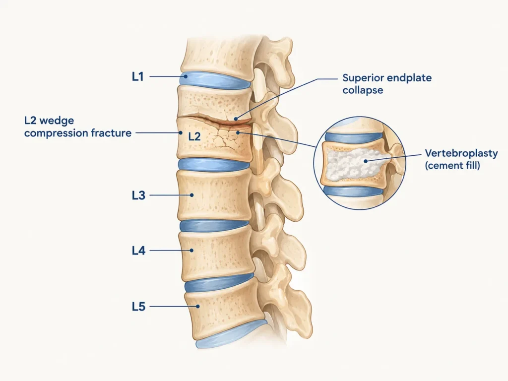
Three Days of Back Pain. Then He Couldn’t Walk. His Vertebra Had Fractured.
At a Glance
| Patient | 59-year-old male |
| Complaint | Severe lower back pain for 3 days; difficulty walking and standing |
| Diagnosis | L2 vertebral body compression fracture with superior endplate collapse |
| Procedure | L2 vertebroplasty |
| Outcome | Fracture stabilised; vertebroplasty changes confirmed on post-operative imaging |
A 59-year-old man came in with three days of severe lower back pain that had been steadily worsening. Walking had become difficult. Standing had become a struggle. He had been hoping it would settle, but it had not.
At 59, a vertebral fracture is not what most people expect, but the scans told a clear story.
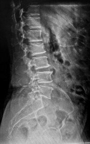
X-ray on arrival showing anterior wedge compression fracture of the L2 vertebral body.
Reach Out for Expert Care
What Did the Scans Show?
Imaging confirmed a compression fracture at L2, the vertebra had been compressed and its upper surface had partially collapsed, causing the pain and difficulty with movement.
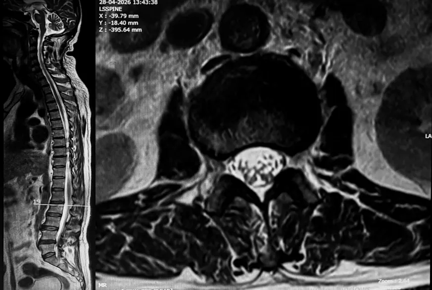
MRI showing L2 compression fracture with partial loss of lumbar lordosis and
disc degeneration across multiple levels.
The MRI showed a compression fracture at L2 with partial loss of the normal lumbar lordosis, the natural inward curve of the lower back had straightened, a sign of pain-related muscle spasm and structural change from the fracture. Disc desiccation changes were also noted across multiple spinal levels, indicating that the discs had lost their normal hydration and cushioning over time, making the spine more vulnerable to injury under load.
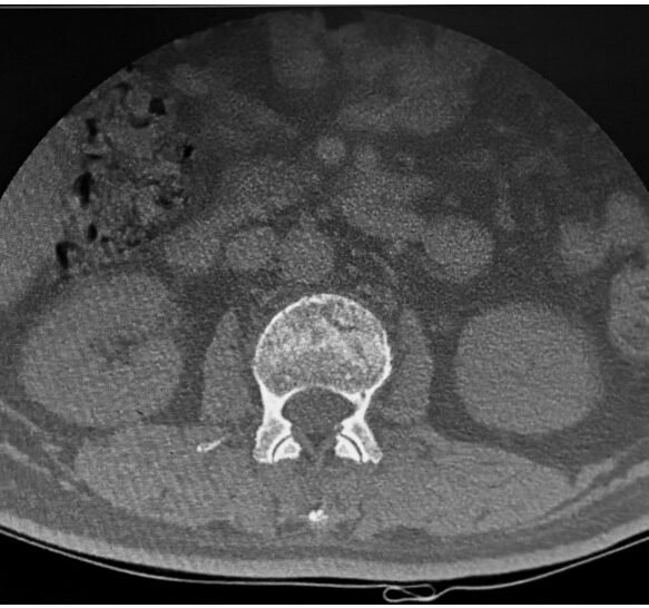
CT scan confirming wedge compression fracture of L2 with superior endplate collapse and
facet joint changes at multiple levels.
The CT scan provided precise bony detail, confirming the wedge compression fracture at L2 with collapse of the superior endplate, the upper surface of the vertebral body. Facet joint arthropathy was noted at multiple lower lumbar levels, and a diffuse disc bulge at L4-L5 was also identified. Together, these findings painted a picture of a spine with underlying degenerative changes, within which the L2 fracture was the acute, painful event requiring immediate treatment.
The Challenge
A compression fracture at L2 in a 59-year-old raises an important question: why did this happen?
Vertebral compression fractures in relatively younger patients, those in their fifties rather than their seventies, are less common and always warrant investigation into bone density and underlying causes. Whether due to osteoporosis, underlying bone weakness, or a specific mechanical event, the fracture itself is both the immediate problem and a signal to look more carefully at the overall bone health.
For the immediate treatment, the goal was clear: stabilise the fractured vertebra, relieve the pain, and allow the patient to return to normal movement as quickly as possible, without the need for a larger open surgical procedure.
Vertebroplasty Procedure: What Was Done?
L2 vertebroplasty, a minimally invasive procedure, was performed to stabilise the fractured vertebra from within, without open surgery.
Vertebroplasty is a percutaneous procedure, performed through the skin, without any surgical incision. Under continuous image guidance, a needle was carefully introduced into the fractured L2 vertebral body through the back. Medical-grade bone cement was then precisely injected into the fractured bone, filling the collapsed spaces within the vertebra and setting firmly to provide immediate structural support.
Think of it as filling a crack in a load-bearing structure so it stops collapsing and holds its shape. Once the cement sets, the vertebra is stabilised, the painful micro-movement within the fracture that causes the severe pain is eliminated, and the bone is supported while it heals.
The procedure was performed without any incision, without general anaesthesia in the conventional surgical sense, and with minimal recovery time compared to open surgery.
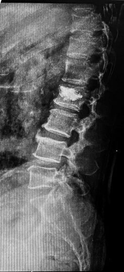
Post-operative X-ray confirming vertebroplasty changes at L2 with cement fill visible,
partial straightening of lumbar spine noted.
Outcome
Post-operative X-ray confirmed successful vertebroplasty at L2, the cement fill was visible within the fractured vertebral body, confirming correct placement and stabilisation of the fracture. Partial straightening of the lumbar spine was noted, consistent with the post-procedural changes expected as the acute pain begins to resolve and muscle spasm gradually settles.
The fractured vertebra was stabilised. The source of the severe pain, the unstable, collapsing fracture, had been addressed through a minimally invasive approach in a 59-year-old patient who had been struggling to walk just days before.
Why Does This Case Matter?
A vertebral fracture is not exclusively an elderly person’s problem. At 59, with underlying disc degeneration and bone changes, a compression fracture can happen, and when it does, it needs prompt diagnosis and treatment.
Vertebral compression fractures are one of the most common causes of acute, severe back pain, yet they are frequently missed or managed with painkillers alone without proper imaging to confirm the diagnosis. Three days of worsening back pain with difficulty walking is not a standard backache. It deserves imaging.
Vertebroplasty has transformed the treatment of compression fractures. What once required open surgery or prolonged bed rest can now be addressed through a needle, under image guidance, with results that allow patients to regain mobility significantly faster than conservative management alone.
The underlying disc degeneration and facet joint changes found in this patient’s scan are also a reminder that spinal health is cumulative. Years of wear, posture, and loading affect the spine long before a fracture makes itself known. Taking spinal symptoms seriously, rather than waiting for the pain to become unbearable, is always the right approach.
Learn more about Minimally Invasive Spine Surgery, Spinal Fixation Surgery, Lumbar Spondylosis and Slip Disc Treatment, or book a consultation for a thorough spinal evaluation.
This case study is published for educational and awareness purposes only. It does not constitute medical advice. Seek prompt medical attention for severe back pain with difficulty walking.

 WhatsApp