
Minimally Invasive Spine Surgery for L1 Vertebral Fracture After a Fall
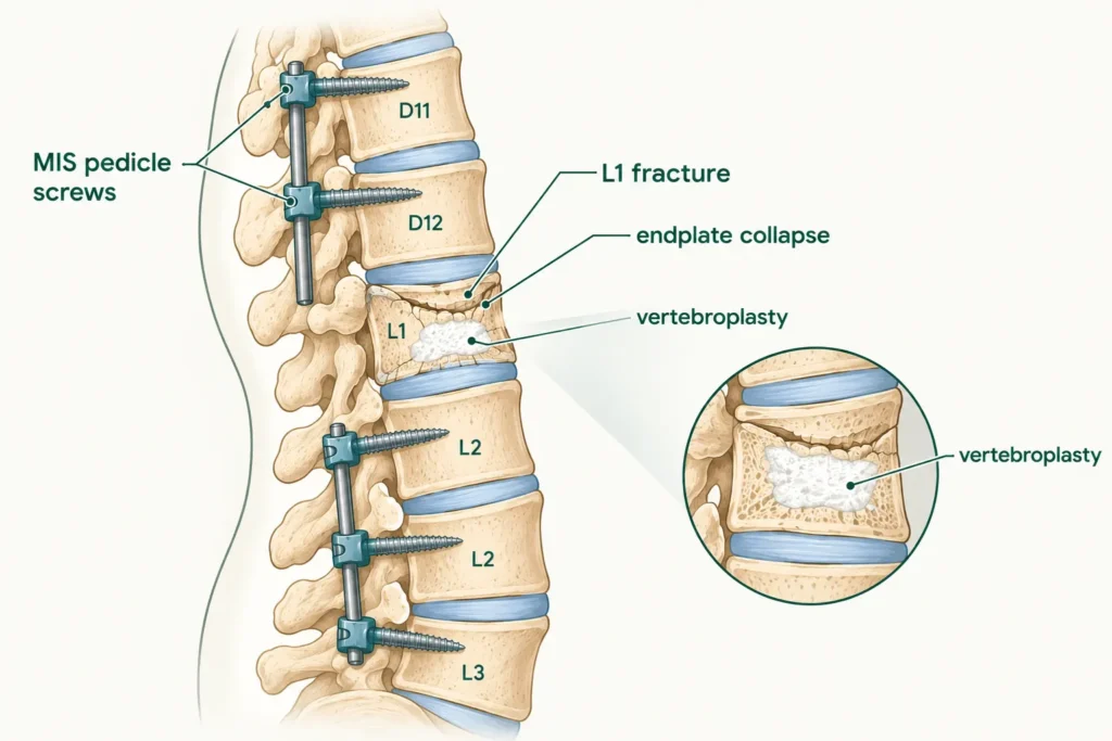
She Fell at Home. By the Time She Reached the Hospital, She Could Barely Move.
At a Glance
| Patient | 77-year-old female |
| Complaint | Severe back pain, urinary hesitancy, restlessness and generalised weakness following a fall at home |
| Diagnosis | L1 vertebral body fracture with superior endplate collapse |
| Procedure | Minimally invasive pedicle screw and rod fixation at D11, D12, L2, L3 with L1 titanium vertebroplasty |
| Outcome | Surgical stabilisation achieved; spine secured with minimally invasive approach |
A 77-year-old woman fell at home that morning. The fall had been triggered by a sudden drop in blood sugar, a hypoglycaemic episode that had left her briefly unable to maintain her balance. When she hit the ground, the impact fractured a vertebra in her lower back.
By the time her family brought her in, she was in severe pain. She was restless and struggling with generalised weakness. She was also experiencing urinary hesitancy, difficulty passing urine, a sign that the fracture was affecting the nerves in the region. She was 77 years old, and her spine needed urgent attention.
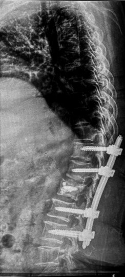
X-ray on arrival showing L1 vertebral fracture following the fall.
Reach Out for Expert Care
What Did the Scan Show?
Imaging confirmed a compression fracture of L1 with the top surface of the vertebra collapsed, and widespread degenerative changes across the spine that made the bone more vulnerable to fracture in the first place.
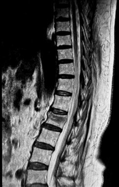
MRI showing L1 vertebral body compression fracture with superior endplate collapse
and degenerative disc changes across multiple levels.
The MRI confirmed a mild wedge compression fracture of the L1 vertebral body, the vertebra had been compressed by the impact of the fall, with the superior endplate collapsing inward.
The MRI also showed degenerative disc changes across multiple levels of the spine, discs that had dried out and lost their normal cushioning, as well as straightening of the normal spinal curve, a sign of long-standing degenerative stress. A mild disc bulge at C3-C4 in the neck was also noted, though this was not the primary concern.
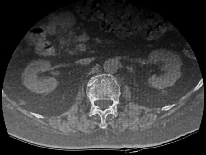
CT scan confirming L1 compression fracture with superior endplate collapse
and disc bulges at multiple lumbar levels.
The CT scan filled in the bony detail, confirming the compression fracture with superior endplate collapse at L1, and showing mild diffuse disc bulges at L2-L3, L3-L4, L4-L5, and L5-S1 levels, all of which were gently pressing on the thecal sac, the protective covering of the spinal nerves. This helped explain the urinary hesitancy and generalised weakness, as the nerve roots in this region were being affected both by the fracture and the surrounding disc pressure.
The Challenge
A fresh vertebral fracture in a 77-year-old with pre-existing spinal degeneration and neurological symptoms is a surgical emergency, but the approach has to be chosen carefully for a patient of this age and frailty.
The L1 fracture was the acute crisis, a collapsed vertebra that needed to be stabilised urgently to prevent further neurological deterioration, but the surrounding spine was already compromised by years of degenerative change, making the bones less able to support standard surgical hardware.
The choice of a minimally invasive approach was deliberate: smaller incisions, less muscle disruption, reduced blood loss, and faster recovery, all critical considerations in a 77-year-old patient presenting with generalised weakness and urinary symptoms.
The decision to use titanium vertebroplasty at L1, rather than the more commonly used bone cement, reflects a considered implant choice. Titanium provides structural support within the fractured vertebra while being fully compatible with future imaging, including MRI, which bone cement can sometimes compromise.
Procedure: What Was Done?
Minimally invasive pedicle screw and rod fixation was performed at D11, D12, L2, and L3, stabilising the spine above and below the fracture, combined with titanium vertebroplasty at L1 to support the fractured vertebra from within.
Minimally Invasive Pedicle Screw and Rod Fixation
Through small incisions, pedicle screws were placed into the vertebrae at D11, D12, L2, and L3, the levels immediately above and below the fractured L1, and connected with rods on either side of the spine. This construct locks the spinal column in correct alignment, transfers the load away from the fractured vertebra, and prevents further collapse or movement at the fracture site. The minimally invasive technique means the muscles alongside the spine are gently moved aside rather than cut, significantly reducing surgical trauma and recovery time.
L1 Titanium Vertebroplasty
At the fractured L1 vertebra itself, a titanium vertebroplasty was performed. A titanium implant was introduced into the collapsed vertebral body to restore its internal structure and provide direct support to the fractured bone from within. Think of it as placing an internal scaffold inside the damaged vertebra to hold its shape and prevent further collapse. Unlike cement vertebroplasty, the titanium implant is permanent, MRI-compatible, and does not carry the risk of cement leakage.
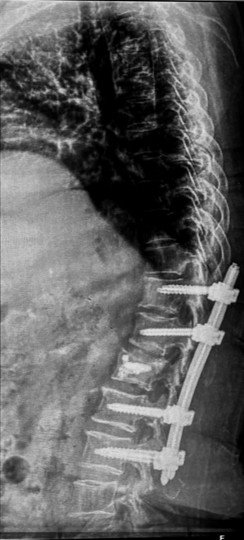
Post-operative X-ray confirming metallic implants and screws correctly positioned
at L1 with fixation at D11, D12, L2, and L3.
Outcome
Post-operative X-ray confirmed the metallic implants and screws were correctly positioned at all treated levels. The spine was stabilised with the vertebroplasty providing internal support to the fractured L1 vertebra and the pedicle screw construct protecting it from further movement.
Surgical stabilisation was successfully achieved in a 77-year-old patient who had presented with an acute fracture, neurological symptoms, and generalised weakness, all managed through a minimally invasive approach that kept surgical trauma to a minimum.
Why Does This Case Matter?
A fall triggered by a medical episode, a sudden drop in blood sugar, a dizzy spell, a moment of weakness, can cause a devastating spinal injury in an elderly person. The fall itself is only half the story.
This case highlights two important awareness points. First, elderly patients with conditions that can cause sudden episodes of weakness or loss of balance, including diabetes, blood pressure fluctuations, or other medical conditions, are at significantly elevated risk of serious injury from a fall. Managing the underlying medical condition is as important as treating the injury that results from it.
Second, vertebral fractures in elderly women are frequently a consequence of osteoporosis, bones that have lost density over years and can fracture under forces that would not harm a younger spine. A fall that might cause a bruise in a 40-year-old can fracture a vertebra in a 77-year-old.
Minimally invasive spine surgery has changed what is possible for elderly patients with acute fractures. Smaller incisions, less blood loss, faster recovery, this approach makes surgical stabilisation accessible to patients who would previously have been considered too frail for open surgery.
If an elderly family member falls, especially one with diabetes, blood pressure issues, or known osteoporosis, a spine evaluation is essential even when the immediate symptoms seem manageable. Back pain after a fall in the elderly is never something to simply rest through.
Learn more about Minimally Invasive Spine Surgery, Spinal Fixation Surgery, Kyphoplasty and Vertebroplasty and Spinal Fracture Treatment or book a consultation
This case study is published for educational and awareness purposes only. It does not constitute medical advice. Seek immediate care for back pain or weakness following a fall in an elderly person.

 WhatsApp