
Combined Burr Hole and MMA Embolisation for Acute on Chronic Subdural Haematoma
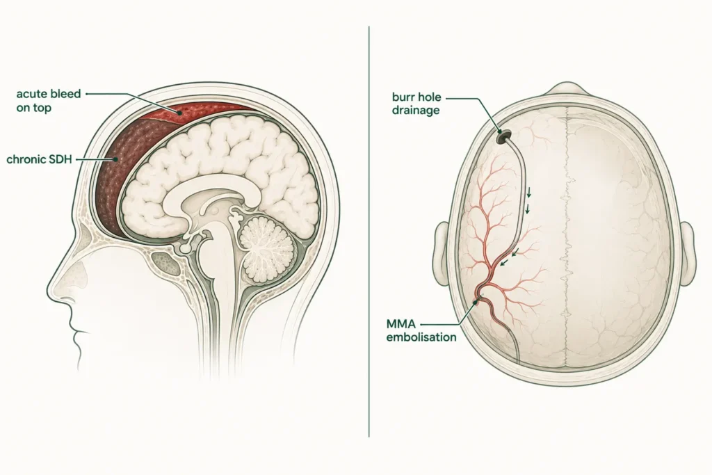
A Fall 10 Days Ago. Then the Weakness Started. Then the Drowsiness. He Was 69.
At a Glance
| Patient | 69-year-old male |
| Complaint | Progressive drowsiness and right-sided weakness developing 10-12 days after a fall |
| Diagnosis | Left fronto-parieto-temporal acute on chronic subdural haematoma |
| Procedure | Cerebral DSA with left MMA embolisation combined with left burr hole and SDH evacuation |
| Outcome | Clinical improvement in drowsiness and right-sided weakness |
A 69-year-old man had a fall about ten days before coming in. At the time, it had not seemed serious. No fracture. No obvious head injury. He had gone home and tried to recover.
Over the following days, something had been quietly changing. He was becoming increasingly drowsy, harder to wake, harder to keep alert. And weakness had been developing on the right side of his body, progressing steadily since the fall.
By the time his family brought him in, the connection between the fall and his deteriorating condition was clear. A brain scan confirmed what had been happening inside his skull for the past ten days.
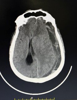
CT scan showing large left-sided subdural haematoma, mixed density collection over the fronto-parieto-temporal region,
indicating acute bleeding on top of a chronic collection.
Reach Out for Expert Care
What Did the Scan Show?
The CT scan revealed a large blood collection covering a wide area of the left side of the brain, old blood with fresh bleeding layered on top, pressing on the brain tissue responsible for movement on the right side of the body.
The NCCT confirmed an extra-axial concavo-convex mixed-density collection along the left fronto-parieto-temporal convexity, a subdural haematoma covering the front, side, and upper regions of the left brain hemisphere.
The mixed density indicated both old and fresh blood, an acute on chronic SDH: a chronic collection that had been present before the fall, with fresh bleeding added on top by the impact.
This explained the timeline precisely. The chronic collection had likely been building slowly before the fall, possibly without causing noticeable symptoms. The fall had triggered fresh bleeding into the existing collection, adding volume, increasing pressure, and producing the progressive drowsiness and right-sided weakness that had developed over the ten days since.
The Challenge
This was a large, mixed collection covering a significant area of the brain, and it needed more than one approach to address it effectively.
In smaller or purely chronic SDH collections, MMA embolisation alone can be sufficient, blocking the artery feeding the haematoma membrane and allowing the body to gradually reabsorb the collection, but when the collection is large, with significant fresh blood and active pressure on the brain, waiting for natural reabsorption alone may not be fast enough to prevent further neurological deterioration.
In this case, the size of the collection and the patient’s progressive symptoms, worsening drowsiness and developing weakness, called for a combined approach: immediate drainage of the haematoma through a burr hole to relieve the pressure quickly, combined with MMA embolisation to cut off the blood supply feeding the membrane and prevent reaccumulation.
MMA embolisation with left burr hole drainage procedure: What Was Done?
A combined procedure was performed: left MMA embolisation to address the root cause, and left burr hole drainage to immediately relieve the pressure, both done together for the best possible outcome.
Left MMA Embolisation
A cerebral DSA, a detailed angiogram of the brain’s blood supply, was performed first to map the left Middle Meningeal Artery and confirm its supply to the haematoma membrane.
The left MMA was then selectively embolised: the artery was blocked from within using the endovascular approach, cutting off the blood supply that had been feeding and maintaining the haematoma.
This addresses the root cause, without it, the collection tends to reaccumulate even after drainage.
Left Burr Hole and SDH Evacuation
Simultaneously, a burr hole, a small opening in the skull, was made on the left side to allow direct drainage of the haematoma. A drainage catheter was placed to evacuate the blood collection and relieve the pressure on the brain immediately. This step rapidly reduces the compression on the brain tissue, giving the neurological symptoms, the drowsiness and weakness, the best chance of improving quickly.
Together, the two procedures work as a team: the burr hole relieves the immediate pressure, and the MMA embolisation ensures the problem does not come back.
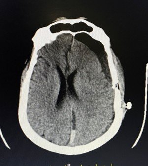
Post-operative CT scan showing left parietal burr hole changes with drainage catheter in situ,
residual collection present as expected in the early post-operative period.
Outcome
Following the combined procedure, the patient showed clinical improvement, the drowsiness began to lift and the right-sided weakness improved. The post-operative CT showed the burr hole changes and drainage catheter in the correct position, with a residual collection visible, which is entirely expected in the early post-operative period.
After SDH drainage, some residual collection always remains initially; this gradually resolves as the brain re-expands and the embolised MMA can no longer feed the membrane.
The combination of immediate drainage and permanent arterial blockade gives this approach its strength, fast relief now, and reduced risk of recurrence later.
Why This Case Matters?
A fall in a person in their sixties or seventies should always be taken seriously, even when it does not seem severe at the time.
One of the most important lessons from this case is the delayed presentation. The fall happened ten to twelve days before the patient came in. During those ten days, a blood collection was slowly growing inside his skull, building pressure, compressing brain tissue, and gradually producing symptoms that could easily have been mistaken for something else. Drowsiness in an elderly person. Weakness on one side. Both dismissed, perhaps, as tiredness or a consequence of the fall itself.
This is the danger of acute on chronic subdural haematoma, the symptoms develop slowly and are easy to miss or misattribute. By the time the connection is made, the collection can be large enough to cause significant neurological damage if not treated promptly.
The second lesson is about surgical planning. Not every SDH is the same. Some can be managed with embolisation alone. Others need drainage alone. Some, like this case, need both, and the decision of which approach to use, and how to combine them, requires experience and careful judgement.
If someone you know has had a fall, even a seemingly minor one, and develops progressive drowsiness, confusion, or weakness in the days that follow, seek immediate medical evaluation. A CT scan can identify the problem in minutes.
Learn more about Subdural Haematoma Treatment, Middle Meningeal Artery Embolisation, Brain Stroke Treatment and Neurointerventional Procedures or book a consultation
This case study is published for educational and awareness purposes only. It does not constitute medical advice. Seek immediate emergency care for progressive drowsiness or weakness following a fall.

 WhatsApp