
Middle Meningeal Artery Embolisation for Chronic Subdural Haematoma
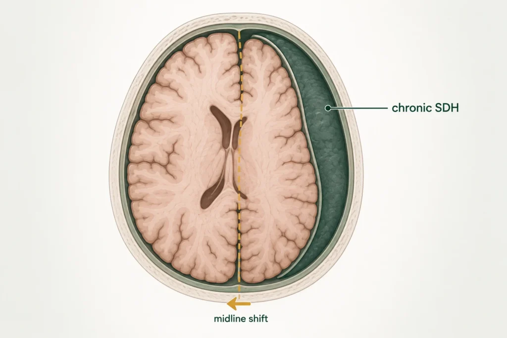
His Family Thought It Was Dementia. It Was a Bleed on the Brain, and It Was Treatable.
At a Glance
| Patient | 83-year-old male |
| Complaint | Forgetfulness and difficulty finding words for 6 months |
| Diagnosis | Left hemispheric chronic subdural haematoma with minimal right frontal subacute SDH |
| Procedure | Digital Subtraction Angiography followed by left Middle Meningeal Artery embolisation |
| Hospital stay | 8 days |
| Outcome | Immediate improvement post-procedure; significantly more oriented at 10-day OPD follow-up |
An 83-year-old man was brought in by his family with a six-month history of increasing forgetfulness and difficulty finding words. He was struggling to retrieve long-term memories, names, events, and conversations. His family had been quietly assuming it was dementia. It was not.
There was no history of a fall. No head injury. No blood thinners. Nothing obvious to explain why an otherwise stable elderly man had been slowly losing his clarity of thought.
The answer was hiding inside his skull.
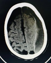
Fig: CT scan showing left hemispheric chronic subdural haematoma,
a slow accumulation of old blood between the brain and its outer covering
Reach Out for Expert Care
What Did the Scan Show?
Imaging revealed a chronic blood collection sitting over the left side of the brain, slowly compressing it, and quietly stealing his memory.
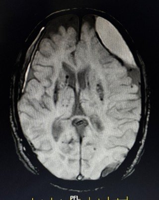
Fig: MRI brain showing left hemispheric chronic subdural haematoma with right frontal subacute SDH,
chronic lacunar infarcts and ischaemic changes
The CT scan and MRI both confirmed a chronic subdural haematoma, an old collection of blood that had accumulated in the space between the brain and its outer protective covering on the left side.
Unlike an acute bleed that happens suddenly after a head injury, a chronic SDH develops slowly over weeks to months, often without any obvious triggering event. The blood gradually accumulates, and the brain, compressed slowly rather than suddenly, adapts until it can no longer compensate.
On the right side, a smaller subacute SDH was also noted in the right frontal region. The MRI additionally showed chronic lacunar infarcts and non-specific ischaemic leukoencephalopathy, changes consistent with his age and background of controlled hypertension, but not the primary cause of his current symptoms.
The left-sided chronic SDH was the culprit. Six months of slowly worsening pressure on the left hemisphere of the brain, the side responsible for language and memory, had been quietly eroding his ability to think, speak, and remember.
The Challenge
In an 83-year-old with a chronic SDH, the decision of how to treat is as important as the decision to treat.
The traditional approach to a symptomatic subdural haematoma is surgical drainage, making a small hole in the skull to drain the collected blood. In younger, healthier patients, this is a straightforward and effective procedure.
In an 83-year-old patient, open surgical intervention carries significantly higher risk. And in a chronic SDH, where the blood has been accumulating over months and has begun to organise, simply draining it does not address the underlying reason it keeps reaccumulating. The middle meningeal artery, a blood vessel that supplies the outer membrane of the brain, is the feeding source of chronic SDHs. As long as it continues to supply blood to the haematoma membrane, the collection tends to recur even after drainage.
The decision was made to treat this with a minimally invasive endovascular approach: Middle Meningeal Artery embolisation, cutting off the blood supply to the haematoma membrane from within the blood vessel itself, without any incision on the skull.
Middle Meningeal Artery Embolisation Procedure: What Was Done?
Dr. Vikas Gupta performed Digital Subtraction Angiography followed by left Middle Meningeal Artery embolisation, a minimally invasive procedure performed entirely through the blood vessels, without open brain surgery.
- A catheter was guided through the blood vessels under continuous X-ray image guidance to the left Middle Meningeal Artery, the artery feeding the outer membrane of the brain where the haematoma had formed. The angiogram confirmed the vascular supply to the haematoma membrane.
- The artery was then selectively embolised.
- Small particles were delivered through the catheter to block the blood supply feeding the chronic haematoma. Without this blood supply, the membrane that had been maintaining and reaccumulating the haematoma can no longer do so, allowing the body to gradually reabsorb the old blood collection over the weeks that follow.
The right frontal subacute SDH was managed conservatively, monitored without intervention, as it was small and not causing significant symptoms.
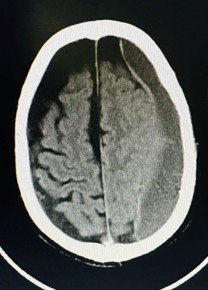
Fig: Post-operative CT scan confirming successful embolisation with changes in the haematoma
consistent with early reabsorption
MMA Embolisation Outcome
Immediately following the procedure, the patient showed early signs of improvement. In chronic SDH cases, the resolution of the haematoma is a gradual process; the brain needs time to reabsorb the old blood collection after the feeding artery has been blocked.
When he returned to the OPD ten days later, the change was clear. He was significantly more oriented, more present in conversation, more able to engage, and noticeably clearer in his thinking than when he had first come in. His family noticed the difference.
He was discharged on day 8, with follow-up imaging scheduled to monitor the progressive reabsorption of the haematoma.
Why Does This Case Matter?
Forgetfulness in an elderly person is almost always assumed to be dementia. This case is a reminder that it is not always that simple, and that some causes of memory loss are entirely reversible.
Chronic subdural haematoma is one of the most commonly missed diagnoses in elderly patients. Since it develops slowly, without a dramatic event, and because its symptoms, forgetfulness, confusion, word-finding difficulty, personality change, closely mimic dementia, it often goes undetected for months. A simple CT scan is all it takes to identify it.
This case also highlights an important advance in how chronic SDH is now being treated. Middle Meningeal Artery embolisation is a relatively new but increasingly evidence-backed approach that addresses the root cause of chronic SDH, the persistent blood supply feeding the haematoma membrane, without the risks of open surgery. In elderly patients where surgical risk is high, this approach offers a safe and effective alternative.
The right frontal SDH in this patient resolved conservatively, a reminder that not every finding on a scan requires immediate intervention. Careful clinical judgement determines what needs to be treated and how.
If an elderly family member has been showing gradual memory loss, increasing confusion, or difficulty with words, and there is no clear diagnosis of dementia, a brain scan is a simple and worthwhile next step.
Learn more about Chronic Subdural Haematoma, Middle Meningeal Artery Embolisation, Brain Stroke Treatment and Neurointerventional Procedures by Dr. Vikas Gupta, or book a consultation for a thorough neurological evaluation.
This case study is published for educational and awareness purposes only. It does not constitute medical advice. If an elderly family member is showing signs of cognitive decline, seek prompt medical evaluation.

 WhatsApp