
Decompressive Hemicraniectomy for Sudden Brain Haemorrhage in a Young Patient
She Was 28. No Health Issues. Then Without Warning: She Collapsed.
At a Glance
| Patient | 28-year-old female |
| Complaint | Sudden onset severe headache followed by loss of consciousness |
| Presentation | GCS E1V1M4 on arrival; right pupil mid-dilated sluggish; left pupil non-reactive; left hemiparesis 1/5 |
| Procedure | Right decompressive hemicraniectomy with embolisation |
| Hospital stay | 8 days |
| Outcome | GCS improved to E4VTM6; hemiparesis improved from 1/5 to 3/5 at discharge; further improved to 4/5 at follow-up; tracheostomy removed; cranioplasty planned |
She was 28 years old. No known health conditions. No warning signs. No history of anything that might have predicted what was about to happen.
A sudden, severe headache, and then she was unconscious.
By the time she arrived, she could barely respond. Her pupils were reacting abnormally. The left side of her body had almost no movement. A young woman in the prime of her life was fighting for it.
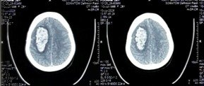
CT scan on arrival showing significant right-sided brain haemorrhage with mass effect and midline shift.
Reach Out for Expert Care
What the Scans Showed
Imaging confirmed a large brain haemorrhage on the right side, with the brain shifting under pressure and the underlying blood vessel abnormality identified on angiography.

CT angiography mapping the blood vessel abnormality responsible for the haemorrhage, essential for planning embolisation.
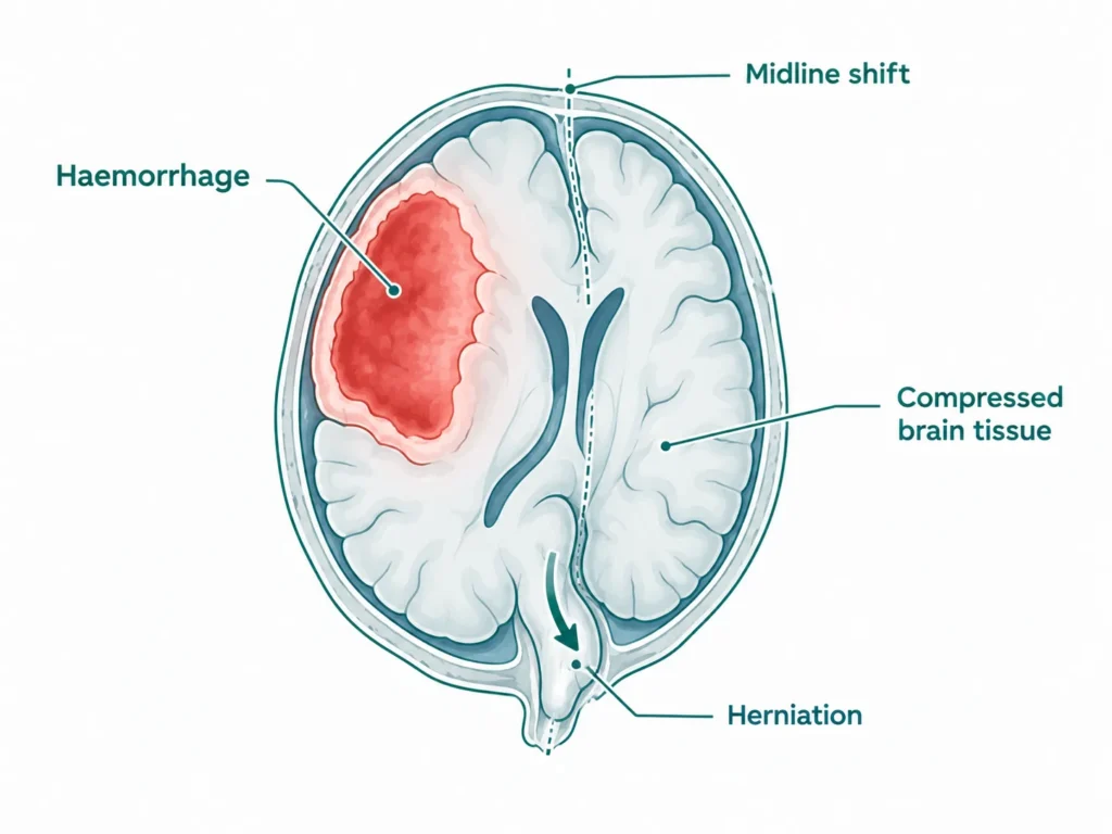
The CT scan confirmed a significant haemorrhage on the right side of the brain with substantial mass effect, the bleeding had created enough pressure to push the brain off its midline, compressing critical structures and accounting for the severe neurological deterioration on arrival.
CT angiography identified the underlying vascular abnormality responsible for the bleed, an abnormal blood vessel that had ruptured, causing the catastrophic haemorrhage. This was not a random event. There had been an underlying structural problem in the blood vessels of the brain, likely present since birth, that had given no warning until this moment.
The Challenge
A 28-year-old with a GCS of E1V1M4, barely responding, pupils reacting abnormally, left side of the body with almost no movement, is in immediate danger of death or permanent severe disability. Every minute matters.
In a haemorrhage of this severity, the brain is under extreme pressure. The bleeding creates a mass that compresses surrounding tissue, cuts off blood supply, and can cause herniation, where the brain is pushed through the base of the skull, a rapidly fatal event if not addressed immediately.
Two things needed to happen simultaneously: the pressure on the brain needed to be relieved urgently through surgery, and the source of the bleed needed to be identified and closed to prevent rebleeding.
Decompressive Hemicraniectomy Procedure: What Was Done?
An emergency right decompressive hemicraniectomy combined with embolisation of the underlying vascular abnormality was performed
Right Decompressive Hemicraniectomy
A large portion of the right side of the skull was temporarily removed, a procedure called decompressive hemicraniectomy. This is not a destructive procedure; it is a life-saving one. When the brain is swelling severely and there is no more room inside the skull, removing part of the skull gives the brain the space it needs to swell safely without being crushed. It is one of the most important emergency neurosurgical procedures available, and in a young patient with a salvageable brain, it can be the difference between survival and death.
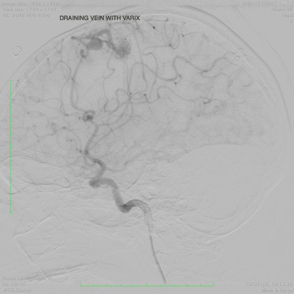
Digital Subtraction Angiography confirming the vascular abnormality and guiding the embolisation procedure.
Embolisation
Following the decompressive surgery, the underlying vascular abnormality identified on angiography was treated with embolisation, a minimally invasive endovascular procedure in which the abnormal blood vessel is blocked from within using a catheter navigated through the blood vessels under image guidance. This closes the source of the bleed, preventing rehaemorrhage while the brain recovers.
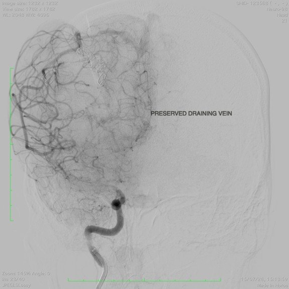
Post-embolisation imaging confirming successful closure of the abnormal blood vessel.
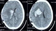
Post-operative CT scan confirming decompression achieved with the hemicraniectomy and resolution of immediate mass effect.
Outcome
The results, given how she had presented, were remarkable.
On arrival, her GCS was E1V1M4, she was barely responsive. By discharge on day 8, her GCS had improved to E4VTM6, she was awake, her eyes opening spontaneously, and responding to commands. The left-sided weakness, which had been 1/5 on arrival, almost no movement at all, had improved to 3/5 by discharge, and further to 4/5 at follow-up, meaning meaningful functional movement had returned.
Her tracheostomy, placed during acute management to protect her airway, was successfully removed. She is planned for cranioplasty, a procedure to replace the portion of skull removed during the hemicraniectomy, as the next step in her recovery.
She came in barely alive. She walked out of the hospital on day 8.
Why This Case Matters
The worst headache of your life is a medical emergency. Always.
A sudden, severe headache, described by patients and neurologists alike as a “thunderclap headache”, that comes on without warning and is unlike anything experienced before is one of the most important warning signs in medicine. It can indicate a brain haemorrhage from a ruptured blood vessel abnormality. In a young, otherwise healthy person, it must be treated as an emergency until proven otherwise.
This case is also a reminder that serious brain conditions do not only affect the elderly. Vascular abnormalities, abnormal blood vessel formations in the brain, can be present from birth and give no warning for decades. When they rupture, they can cause catastrophic haemorrhage in young, healthy people with no prior symptoms and no known risk factors.
The outcome in this case, a young woman who arrived with GCS E1V1M4 and left the hospital awake and moving her left arm and leg, reflects what is possible when a neurovascular emergency is recognised and treated immediately by an experienced team.
Time is brain. The faster the intervention, the more of it is saved.
Learn more about Brain Haemorrhage Treatment, Neurointerventional Procedures, Brain AVM Treatment and Emergency Neurosurgery or book a consultation with Dr. Vikas Gupta.
This case study is published for educational and awareness purposes only. It does not constitute medical advice. A sudden severe headache is a medical emergency, call for emergency care immediately.

 WhatsApp