
Disc Surgery for Leg Weakness and Back Pain in Osteoporosis
Reach Out for Expert Care
She Could Barely Walk. Her Spine Was Collapsing, and Her Bones Were Too Weak to Fight Back.
At a Glance
| Patient | 74-year-old female |
| Complaint | Severe lower back pain; weakness in both legs; difficulty walking; numbness in right foot |
| Diagnosis | L3-L4 disc prolapse; L1 endplate fracture; severe osteoporosis |
| Procedure | L3-L4 discectomy with pedicle screw and rod fixation at L3-L4 |
| Hospital stay | 10 days |
| Outcome | Pain relieved; leg weakness improved; numbness resolved; walking restored |
A 74-year-old woman came in unable to walk properly. Her lower back pain was severe, and weakness had spread into both her legs, with numbness settling into her right foot. Simple things like standing up and moving around had become a significant struggle.
What made this case more than a straightforward disc problem was what lay beneath: severe osteoporosis, bones so weakened that they could barely hold the spine’s own weight, and a fracture at L1 that had gone unaddressed. Her spine was under pressure from multiple directions at once.
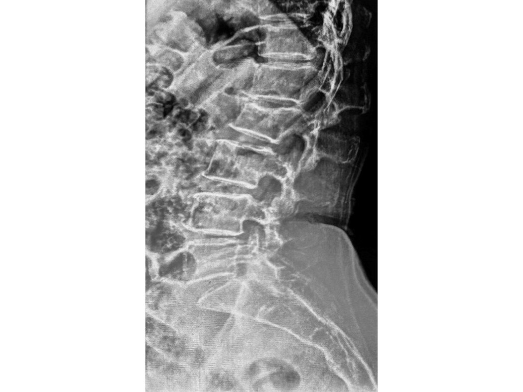
X-ray on arrival showing reduced disc space at L1-L2 and L4-L5, with partial straightening of the lumbar spine.
What Did the Scan Show?
Imaging revealed a severely compressed spinal canal at L3-L4, with the nerve roots on both sides being squeezed, explaining the weakness and numbness in her legs.
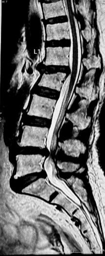
MRI showing severe disc bulge at L3-L4 with canal narrowing and bilateral nerve root compression.
The MRI showed a significant disc prolapse at L3-L4, the disc had bulged outward with reduced height, thickening of the ligamentum flavum, and facet joint arthropathy all contributing to a severely narrowed spinal canal.
The thecal sac, the protective sheath surrounding the spinal cord and nerve roots, had been compressed to approximately 5mm in diameter at this level. Both the traversing and exiting nerve roots on either side were being squeezed. This directly explained the weakness in both legs and the numbness in the right foot.
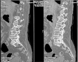
CT scan showing reduced disc height at L5-S1 with endplate irregularities and sclerosis, signs of long-standing degenerative change.
The CT scan confirmed additional degenerative changes at L5-S1, reduced disc height, endplate irregularities, and sclerosis, painting a picture of a spine that had been under significant stress for many years.
Combined with a DEXA scan showing severe osteoporosis with a T-score of -3.8, the full picture was clear: this was a fragile spine in crisis.
The Challenge
Operating on a 74-year-old with severe osteoporosis and multi-level degeneration is not routine. Every decision, from implant choice to surgical approach, had to account for bones that were significantly weaker than normal.
The primary problem was the L3-L4 disc prolapse compressing the nerve roots. But the osteoporosis complicated everything. Standard pedicle screws may not hold adequately in severely osteoporotic bone, risking implant failure after surgery. The L1 endplate fracture added another layer of instability to an already compromised spine.
The surgical plan had to achieve two things: decompress the nerves causing the weakness and numbness, and stabilise the spine in bone that could not be treated like a younger patient’s.
Procedure of Disectomy: What Was Done?
Dr. Vikas Gupta performed L3-L4 discectomy with pedicle screw and rod fixation, removing the disc compressing the nerves and stabilising the spine in the same procedure.
The prolapsed disc at L3-L4 was carefully removed, taking the pressure off the compressed nerve roots on both sides. This is called a discectomy: a precise removal of the disc material that has pushed out of its normal position and is pressing on the nerves.
Pedicle screws and rods were then placed at L3-L4 to stabilise the operated level and prevent further instability in an already degenerative spine.
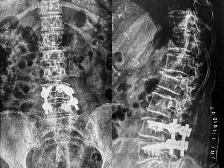
Post-operative X-ray confirming metallic rod and screw fixation correctly positioned at L3-L4.
Both intraoperative and post-operative imaging confirmed the implants were correctly placed and the construct was well positioned across the operated level.
Outcome
Following spine surgery in Delhi, the patient experienced significant relief. The weakness in both legs improved. The numbness in her right foot resolved. She was able to walk again, a meaningful restoration of function for a 74-year-old who had arrived barely able to move.
She was discharged on day 10 with guidance on mobilisation and follow-up care.
Why Does This Case Matter?
Leg weakness and numbness in elderly patients are often attributed to age or general frailty. This case is a reminder that these symptoms, particularly when progressive, can have a clear, treatable spinal cause.
Lumbar disc prolapse does not only affect younger people. In older adults, the combination of disc degeneration, spinal canal narrowing, and osteoporosis can create a perfect storm, where the nerves are being compressed from multiple directions simultaneously. The result is weakness, numbness, and loss of the ability to walk independently.
What this case also illustrates is the importance of specialist surgical planning in elderly patients with osteoporosis. Surgery in this group is not impossible, but it requires a different approach, different implant considerations, and a thorough understanding of the spine’s fragility before a single screw is placed.
If an elderly family member is developing weakness in the legs, difficulty walking, or numbness that is getting progressively worse, do not assume it is just age. Get a proper spinal evaluation.
Learn more about Slip Disc Treatment in Delhi, Microdiscectomy Surgery, Spinal Fixation Surgery, and Lumbar Spondylosis, or book a consultation with Dr. Vikas Gupta for a thorough spinal evaluation.
This case study is published for educational and awareness purposes only. It does not constitute medical advice.

 WhatsApp